Summary
Choledocholithiasis, or stones in the bile duct, is a condition closely related to gallstones. It occurs when gallstones migrate from the gallbladder and block the bile ducts that lead to the small intestine. The most commonly affected areas include the cystic duct, common bile duct, and common hepatic duct.
The gallbladder, located beneath the liver, serves as a reservoir for bile before it flows into the small intestine. Bile is a digestive fluid that helps break down fats in the food we eat. Gallstones form when bile contains an excess of cholesterol or bilirubin, a substance produced when the liver breaks down old red blood cells.
Symptoms of choledocholithiasis can include pain in the upper right or central part of the abdomen, nausea, vomiting, yellowing of the skin and eyes (jaundice), and pale-colored stools.
There are several treatment options for this condition, including surgical procedures to remove the stones or, in more severe cases, removal of the gallbladder itself.
Table of Contents
Symptoms of Choledocholithiasis
In many cases, choledocholithiasis (stones in the bile duct) does not produce noticeable symptoms unless the stones block the bile ducts. When this happens, symptoms can range from mild discomfort to severe pain and other complications. Here are some common symptoms to watch for:
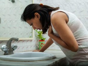
- Pain in the upper right or upper central abdomen. This pain can be intense and continuous or may come in waves, lasting for around 30 minutes or more. It is often felt in the upper right side of the abdomen but can also occur in the center.
- Yellowing of the skin and eyes (jaundice). A blockage in the bile duct can cause bile to back up into the liver, leading to jaundice. This results in yellowing of the skin and the whites of the eyes.
- Fever. A high temperature may occur if the blockage leads to an infection in the bile ducts or gallbladder, indicating a more serious complication.
- Nausea and vomiting. Digestive distress, including nausea and vomiting, often accompanies the pain, especially after eating fatty foods.
- Loss of appetite. Due to the discomfort and digestive issues caused by the blockage, many people experience a reduced desire to eat.
- Pale or clay-colored stools. When bile flow is blocked, it cannot reach the intestines, causing the stool to lose its normal brown color and appear pale or grayish.
Several factors can increase the risk of developing choledocholithiasis, such as obesity, rapid weight loss, or a high-fat diet. If any of these symptoms occur, particularly jaundice or fever, it is important to seek medical attention to avoid complications like infection or pancreatitis.
Types of Choledocholithiasis
There are two main types of gallstones that can lead to choledocholithiasis, each formed by different substances within the bile. Understanding these types is important for diagnosing and treating the condition effectively:
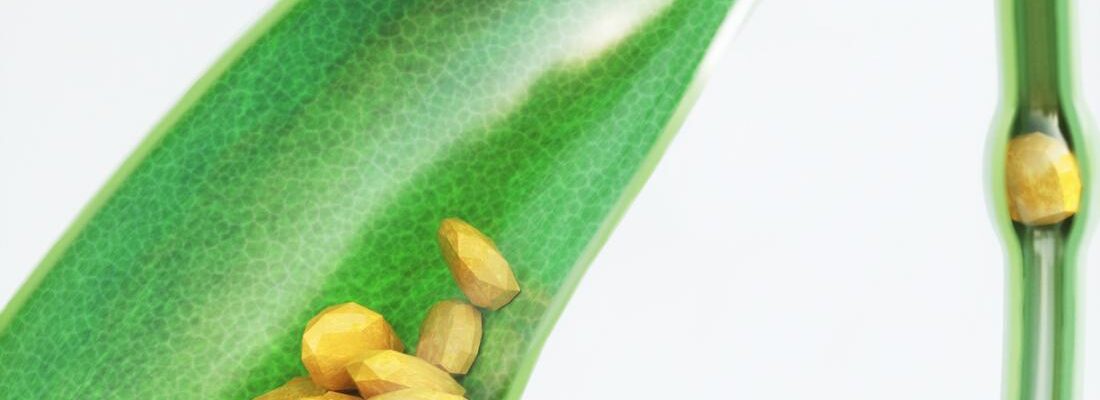
- Cholesterol Stones. These are the most common type of gallstones. They form when there is too much cholesterol in the bile, which can’t be dissolved completely. Over time, this excess cholesterol crystallizes into solid stones. Cholesterol stones are usually yellowish in color and often develop when there is an imbalance in the substances that break down fats in the bile.
- Pigment Stones (Bilirubin Stones). These stones form when there is too much bilirubin in the bile. Bilirubin is a byproduct of the breakdown of red blood cells, which is processed by the liver. Certain conditions, such as liver disease or infections of the biliary system, can cause an excess of bilirubin, leading to the formation of these darker, black or brown stones.
Both types of stones can migrate from the gallbladder into the bile ducts, where they cause blockages and lead to symptoms of choledocholithiasis. Identifying the type of stone is essential for determining the most appropriate treatment, as different stones may respond differently to medication or surgical intervention.
Diagnostic Procedures for Choledocholithiasis
Diagnosing choledocholithiasis, or the presence of gallstones in the bile ducts, requires a combination of clinical evaluation, imaging studies, and sometimes specialized tests to confirm the condition and assess its severity. Here are the key diagnostic procedures used:
- Ultrasound. This is often the first imaging test performed to detect gallstones. An ultrasound uses sound waves to create images of the gallbladder and bile ducts. While it is excellent for detecting stones in the gallbladder, it may not always identify stones within the bile ducts themselves.
- Endoscopic Ultrasound (EUS). This procedure is more sensitive than a standard ultrasound, especially for detecting smaller stones in the bile ducts. EUS uses a flexible tube with an ultrasound probe on the tip, inserted through the mouth and into the stomach and small intestine, to get detailed images of the bile ducts and surrounding organs.
- Magnetic Resonance Cholangiopancreatography (MRCP). MRCP is a specialized MRI scan that provides high-resolution images of the bile ducts, gallbladder, liver, and pancreas. It is a non-invasive and highly accurate way to identify stones or blockages in the bile ducts.
- Endoscopic Retrograde Cholangiopancreatography (ERCP). ERCP is both a diagnostic and therapeutic procedure. A flexible endoscope is passed through the mouth into the small intestine, where contrast dye is injected into the bile ducts. X-ray images are then taken to detect blockages. If gallstones are found, they can often be removed during the procedure, making ERCP a valuable tool for both diagnosis and treatment.
- CT Scan. A computed tomography (CT) scan may be used to detect complications from choledocholithiasis, such as infection, inflammation, or abscesses. While CT scans are less effective at detecting gallstones themselves, they provide valuable information about the overall condition of the abdomen.
- Blood Tests. Although blood tests cannot directly detect gallstones, they can provide important clues about liver function, bile duct obstruction, or infection. Elevated levels of liver enzymes, bilirubin, or white blood cells may suggest a blockage or inflammation in the bile ducts.
These diagnostic procedures help determine the presence, location, and severity of bile duct stones. Depending on the results, the appropriate treatment plan—whether it involves medication, non-invasive removal techniques, or surgery—can be decided to prevent further complications.
Complications of Untreated Choledocholithiasis
f left untreated, choledocholithiasis (gallstones in the bile ducts) can lead to serious and potentially life-threatening complications. The obstruction of the bile ducts caused by gallstones disrupts the normal flow of bile and digestive enzymes, leading to a range of health issues. Here are some of the most common complications of untreated choledocholithiasis:
- Cholangitis (Infection of the Bile Ducts). One of the most serious complications, cholangitis occurs when a bile duct blockage leads to bacterial infection. Symptoms include fever, chills, jaundice, and severe abdominal pain. If untreated, cholangitis can cause sepsis, a life-threatening infection that spreads throughout the body.
- Acute Pancreatitis. When a gallstone blocks the pancreatic duct, digestive enzymes can become trapped within the pancreas, causing inflammation. This condition, known as pancreatitis, leads to intense abdominal pain, nausea, vomiting, and fever. Acute pancreatitis can range from mild to severe and may require hospitalization.
- Biliary Cirrhosis. Prolonged blockage of the bile ducts can cause bile to accumulate in the liver, leading to liver damage over time. This damage may result in biliary cirrhosis, a condition in which healthy liver tissue is replaced by scar tissue, impairing liver function and potentially leading to liver failure.
- Gallbladder Inflammation (Cholecystitis). If the blockage occurs in the cystic duct (which connects the gallbladder to the common bile duct), it can lead to inflammation of the gallbladder, known as cholecystitis. Symptoms include severe abdominal pain, fever, and nausea. In severe cases, the gallbladder may become infected or even rupture, requiring emergency surgery.
- Bile Duct Stricture. Chronic inflammation or repeated episodes of choledocholithiasis can cause scarring and narrowing of the bile ducts (known as strictures). This can lead to long-term bile flow obstruction and recurrent episodes of infection, pain, and jaundice.
- Hepatic Abscess. In rare cases, untreated choledocholithiasis can lead to the formation of an abscess in the liver, caused by a bacterial infection. A hepatic abscess can cause fever, abdominal pain, and sepsis if not promptly treated.
- Secondary Biliary Cirrhosis. If the bile ducts remain obstructed for a long period, secondary biliary cirrhosis can develop. This condition occurs when the liver becomes progressively damaged due to prolonged bile buildup, potentially leading to liver failure.
Untreated choledocholithiasis can quickly progress to more severe health issues, many of which are life-threatening. Early detection and timely treatment, including the removal of the stones and restoration of bile flow, are critical in preventing these complications.
Causes of Choledocholithiasis
Gallstones in the bile ducts, or choledocholithiasis, develop due to a variety of factors that affect the composition and flow of bile. These factors lead to the formation of stones, either from cholesterol or bilirubin, which can block the bile ducts and cause complications. Here are the main causes:
- Cholesterol buildup in bile. When the bile contains more cholesterol than it can dissolve, excess cholesterol may crystallize and form gallstones. This is one of the most common causes of stone formation.
- Excess bilirubin in bile. Bilirubin is a substance produced when the liver breaks down old red blood cells. Certain conditions, such as liver disease or blood disorders, can lead to excess bilirubin in the bile, which can form pigment stones that are darker in color.
- Lack of bile salts. Bile salts are crucial for breaking down fats in the digestive process. When the bile contains insufficient bile salts, it becomes more concentrated, increasing the likelihood of gallstone formation.
- Incomplete emptying of bile. If the gallbladder does not fully empty its bile, the remaining bile becomes concentrated and may cause particles to cluster and form stones.
- Infection in the bile ducts or liver. Bacterial infections in the bile ducts or liver can alter the chemical balance of bile, leading to the formation of gallstones and increasing the risk of bile duct blockages.
- Cirrhosis. Cirrhosis, a condition in which the liver is severely scarred and damaged, can alter bile production and flow, making it more likely for gallstones to develop.
- Hereditary blood disorders. Some inherited conditions, such as hemolytic anemia, cause the body to break down red blood cells at a faster rate, resulting in higher bilirubin levels. This excess bilirubin can lead to the formation of pigment stones in the bile ducts.
These factors, whether related to cholesterol, bilirubin, or bile flow, can all contribute to the development of gallstones and the condition known as choledocholithiasis. Identifying the underlying cause is important in preventing recurrence and choosing the appropriate treatment.
Prevention of Choledocholithiasis

Even if someone has had gallstones in the past, there is still a risk of developing new stones, particularly in the bile ducts, even after the gallbladder has been removed. However, there are several strategies to reduce the risk of gallstone formation and prevent choledocholithiasis. These include lifestyle changes and dietary adjustments aimed at maintaining a healthy bile flow and reducing cholesterol buildup.
Here are effective ways to prevent choledocholithiasis:
- Regular exercise. Physical activity plays a significant role in preventing the formation of gallstones. Engaging in at least 30 minutes of exercise daily, or five times a week, helps maintain a healthy weight and improves the body’s ability to metabolize fats, reducing the risk of gallstones.
- Eating high-fiber foods. A diet rich in fiber aids digestion and helps lower cholesterol levels in the digestive system. Foods like fruits, vegetables, whole grains, and legumes can promote healthy bile production and reduce the likelihood of stone formation.
- Limiting saturated fats. Saturated fats, found in fatty meats, processed foods, and full-fat dairy products, are more difficult for the body to break down. Reducing intake of these fats can prevent them from accumulating in the bile and forming stones.
- Staying hydrated. Drinking plenty of fluids, especially water, supports healthy digestion and prevents bile from becoming too concentrated. Staying well-hydrated also promotes efficient metabolism of food, reducing the risk of gallstone formation.
By adopting these preventive measures, individuals can significantly lower their chances of developing choledocholithiasis and improve their overall digestive health. Regular exercise, a fiber-rich diet, and proper hydration are key factors in maintaining a healthy bile flow and preventing complications related to gallstones.
Risk Factors for Choledocholithiasis

Several factors can increase the likelihood of developing gallstones, which may later lead to choledocholithiasis (stones in the bile ducts). These risk factors involve a combination of biological, genetic, and lifestyle-related elements. Understanding these risks can help with prevention and early detection of the condition.
Here are the common risk factors for choledocholithiasis:
- Female gender and elevated estrogen levels. Women, particularly those with higher estrogen levels, are at greater risk of gallstone formation, which can lead to bile duct stones.
- Pregnancy. Hormonal changes during pregnancy can increase the risk of developing gallstones, which may block the bile ducts.
- Use of hormone replacement therapy or contraceptives. These medications can raise estrogen levels, which may slow down bile flow and increase the risk of stone formation.
- Age over 40. The risk of gallstone and bile duct stone development increases with age, particularly after 40.
- Family history of gallstones. Genetics play a significant role, and having a family member with gallstones increases your own risk.
- Obesity. Excess body fat raises cholesterol levels, increasing the chance of gallstones forming in the bile and leading to potential blockages.
- Rapid weight loss. Losing weight too quickly can cause the liver to produce more cholesterol, which may lead to the development of stones.
- High-calorie, high-carbohydrate diet. Diets rich in calories and refined carbohydrates increase the likelihood of gallstone formation.
- Low-fiber diet. Insufficient fiber in the diet can lead to digestive issues and increase the risk of stone formation.
- Native American or Mexican-American ethnicity. Certain ethnic groups have a higher predisposition to developing gallstones and bile duct stones due to genetic factors.
- Digestive disorders, such as Crohn’s disease. Conditions affecting the digestive tract can interfere with bile metabolism and increase the risk of gallstones.
- Diabetes and metabolic syndrome. These conditions elevate triglyceride levels in the blood, increasing the risk of stone formation.
- Cirrhosis or bile duct infections. Liver diseases, including cirrhosis, and infections in the bile ducts can cause bile to become imbalanced, leading to stone formation.
- Hemolytic anemia, such as sickle cell disease. Blood disorders that cause rapid breakdown of red blood cells can increase bilirubin levels, leading to pigment stone formation.
- Previous gallstones. A history of gallstones increases the likelihood of stones recurring in the bile ducts.
- Previous gallbladder removal. Even after gallbladder removal, there is still a chance of developing stones in the bile ducts.
By recognizing these risk factors, individuals can take preventive steps, such as maintaining a healthy diet, managing weight, and addressing underlying health conditions, to reduce the likelihood of developing choledocholithiasis.
Choledocholithiasis FAQs
Here are some frequently asked questions (FAQs) about choledocholithiasis, or bile duct stones, to help you better understand this condition:
- What is choledocholithiasis?
Choledocholithiasis is the presence of gallstones in the bile ducts. These stones usually form in the gallbladder and then move into the bile ducts, where they can cause blockages and disrupt the flow of bile to the small intestine. - What causes choledocholithiasis?
Choledocholithiasis occurs when gallstones form due to imbalances in bile components, such as cholesterol or bilirubin. These stones can then migrate from the gallbladder to the bile ducts, where they may cause a blockage. Factors like obesity, rapid weight loss, and certain medical conditions (e.g., diabetes, liver disease) increase the risk of developing bile duct stones. - What are the symptoms of choledocholithiasis?
Common symptoms include upper right or central abdominal pain, jaundice (yellowing of the skin and eyes), nausea, vomiting, fever, and pale or clay-colored stools. In severe cases, it can cause life-threatening infections or inflammation of the pancreas (pancreatitis). - How is choledocholithiasis diagnosed?
Diagnosis typically involves imaging tests such as ultrasound, endoscopic ultrasound (EUS), magnetic resonance cholangiopancreatography (MRCP), or endoscopic retrograde cholangiopancreatography (ERCP). Blood tests may also be used to check for liver function or signs of infection. - What is the treatment for choledocholithiasis?
The primary treatment is the removal of the stones. This is often done through ERCP, where an endoscope is used to locate and extract the stones. In some cases, surgery may be required to remove the gallbladder (cholecystectomy) or address complications. Antibiotics may also be prescribed if an infection is present. - Can choledocholithiasis go away on its own?
It is rare for bile duct stones to pass on their own. Most cases require medical intervention, especially if symptoms are present. Without treatment, choledocholithiasis can lead to serious complications, including infections or pancreatitis. - What are the complications of untreated choledocholithiasis?
Untreated choledocholithiasis can lead to serious conditions like cholangitis (infection of the bile ducts), pancreatitis (inflammation of the pancreas), biliary cirrhosis (liver damage), and bile duct strictures. In severe cases, it can cause sepsis, a life-threatening infection that spreads throughout the body. - Can you develop choledocholithiasis after gallbladder removal?
Yes, even after gallbladder removal (cholecystectomy), it is still possible to develop bile duct stones. This can happen if stones form in the liver or if residual stones from the gallbladder migrate to the bile ducts after surgery. - How can I prevent choledocholithiasis?
Prevention strategies include maintaining a healthy weight, exercising regularly, eating a diet high in fiber and low in saturated fats, and avoiding rapid weight loss. Managing underlying health conditions like diabetes or liver disease also helps reduce the risk of stone formation. - Is choledocholithiasis a life-threatening condition?
Choledocholithiasis can become life-threatening if not treated, especially if it leads to complications such as cholangitis, pancreatitis, or sepsis. Timely diagnosis and treatment are essential to prevent severe outcomes.
These FAQs provide key information about choledocholithiasis, including its causes, symptoms, diagnosis, and treatment. If you experience symptoms of bile duct stones, it is important to seek medical attention promptly to avoid serious complications.